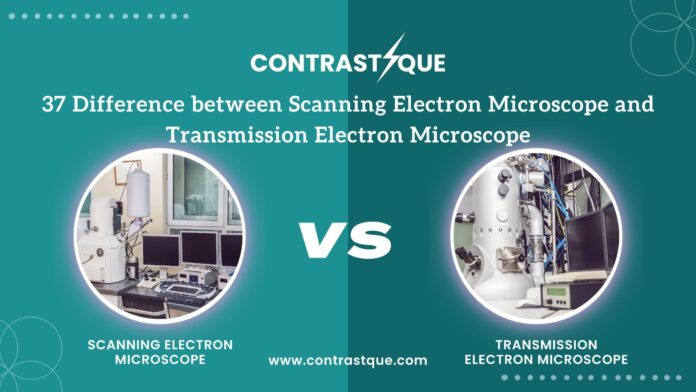
Introduction to electron microscopes
Are you ready to dive into the fascinating world of electron microscopy? Get ready to uncover the intricate details and differences between two powerful imaging tools – the Scanning Electron Microscope (SEM) and Transmission Electron Microscope (TEM). Join us on a journey through 37 key distinctions that set these groundbreaking technologies apart, from imaging techniques to applications and beyond. Let’s unlock the secrets of SEM and TEM together!
Scanning Electron Microscope (SEM): Definition and Functionality
Are you ready to dive into the world of scanning electron microscopes (SEM)? Let’s explore the fascinating realm of SEM technology and its functionalities.
SEM is a powerful imaging tool that uses focused beams of electrons to create high-resolution images of samples. Unlike optical microscopes, SEM can achieve magnifications up to 100,000 times, revealing intricate details at the nanoscale level.
One key feature of SEM is its ability to produce 3D images through a process called electron tomography. This technique allows researchers to visualize the internal structure of specimens in unprecedented detail.
In addition to imaging capabilities, SEM can also analyze elemental composition using energy-dispersive X-ray spectroscopy (EDS). This provides valuable insights into the chemical makeup of materials being studied.
SEM plays a crucial role in various scientific fields such as material science, biology, and geology. Its versatility and high resolution make it an indispensable tool for researchers seeking to understand the microscopic world around us.
Transmission Electron Microscope (TEM): Definition and Functionality
The Transmission Electron Microscope (TEM) is a powerful tool used in the field of microscopy to observe ultra-thin samples at high resolution. Unlike its counterpart, the Scanning Electron Microscope, TEM operates by transmitting electrons through a specimen rather than scanning its surface. This allows for detailed imaging of internal structures and atomic level features.
In TEM, electron beams pass through the sample creating an image on a fluorescent screen or digital detector. The resulting images provide intricate details about the sample’s composition and structure. With its ability to achieve magnifications up to 2 million times, TEM is essential in various scientific disciplines ranging from materials science to biology.
By utilizing electromagnetic lenses and advanced imaging techniques, Transmission Electron Microscopes offer unparalleled clarity and precision in visualizing nanoscale objects. Researchers rely on TEM for studying nanoparticles, viruses, crystals, and even individual atoms with remarkable accuracy.
Let’s Explore 37 Difference between scanning electron microscope and transmission electron microscope
Let’s delve into the intricate world of electron microscopy and uncover the nuances that distinguish scanning electron microscopes (SEMs) from transmission electron microscopes (TEMs). These powerful tools offer unique capabilities for imaging at the nanoscale, revolutionizing scientific research across various fields.
In terms of imaging techniques, SEMs utilize a focused beam of electrons to scan surfaces, producing detailed 3D images. On the other hand, TEMs transmit electrons through thin samples to create high-resolution 2D images of internal structures.
When it comes to sample preparation, SEMs typically require coating samples with a conductive material to enhance image quality, while TEMs necessitate ultrathin specimen sections for transmission through the sample.
Magnification and resolution differ between SEM and TEM; SEM provides higher magnification for surface details, whereas TEM offers superior resolution for internal structures due to shorter wavelengths of transmitted electrons.
These distinctions in functionality highlight the diverse applications of SEM and TEM in materials science, biology, nanotechnology, and more. Whether analyzing nanoparticles or studying cellular organelles with atomic precision, both instruments play a vital role in advancing scientific knowledge.
|
S. No. |
Aspect |
Scanning Electron Microscope (SEM) |
Transmission Electron Microscope (TEM) |
|
1 |
Basic Principle |
Scans a focused electron beam across the surface to create an image |
Transmits electrons through a thin specimen to form an image |
|
2 |
Electron Interaction |
Surface interaction |
Transmission through specimen |
|
3 |
Image Formation |
Secondary electrons emitted from the surface |
Electrons passing through the specimen |
|
4 |
Resolution |
Lower (1-20 nm) |
Higher (0.1-0.5 nm) |
|
5 |
Magnification |
20x to 30,000x |
Up to 1,000,000x |
|
6 |
Sample Preparation |
Less intensive, usually requires conductive coating |
More intensive, requires ultra-thin slicing |
|
7 |
Sample Thickness |
Any thickness |
Very thin (less than 100 nm) |
|
8 |
Image Dimension |
3D surface images |
2D images |
|
9 |
Type of Electrons Used |
Secondary and backscattered electrons |
Transmitted electrons |
|
10 |
Depth of Field |
High |
Low |
|
11 |
Image Contrast |
Topographical and compositional contrast |
Amplitude and phase contrast |
|
12 |
Usage |
Surface structure and composition analysis |
Internal structure analysis |
|
13 |
Operating Voltage |
0.1 to 30 kV |
60 to 300 kV |
|
14 |
Working Environment |
Vacuum |
High vacuum or ultra-high vacuum |
|
15 |
Detectors |
Secondary electron detector, backscatter detector, X-ray detector |
Electron energy loss, diffraction, X-ray detectors |
|
16 |
Imaging Speed |
Faster |
Slower |
|
17 |
Ease of Use |
Easier |
More complex |
|
18 |
Cost |
Generally lower |
Generally higher |
|
19 |
Sample Limitations |
Conductive or coated non-conductive |
Must be electron-transparent |
|
20 |
Elemental Analysis |
Energy-dispersive X-ray spectroscopy (EDS) |
Electron energy loss spectroscopy (EELS) |
|
21 |
Crystallographic Information |
Limited |
Extensive (diffraction patterns) |
|
22 |
Maintenance |
Lower |
Higher |
|
23 |
Beam Damage |
Less likely |
More likely |
|
24 |
Image Artifacts |
Charging effects on non-conductive samples |
Diffraction and thickness artifacts |
|
25 |
Environmental SEM (ESEM) Capability |
Yes |
No |
|
26 |
Biological Sample Imaging |
Limited, requires coating |
Better, with staining |
|
27 |
Phase Information |
Not typically available |
Available through phase contrast |
|
28 |
Surface Detail |
Excellent |
Limited |
|
29 |
In Situ Observation |
Limited |
Possible with special holders |
|
30 |
Resolution in Vacuum |
High |
Very high |
|
31 |
Beam Penetration |
Shallow |
Deep |
|
32 |
Auger Electron Spectroscopy (AES) |
Yes |
No |
|
33 |
Heating and Cooling Stages |
Available |
Available but more complex |
|
34 |
X-ray Microanalysis |
Commonly integrated |
Can be integrated |
|
35 |
STEM Mode |
Not applicable |
Yes (Scanning TEM) |
|
36 |
Focused Ion Beam (FIB) Compatibility |
Yes |
No |
|
37 |
Sample Size |
Can handle larger samples |
Limited to small samples |
Differences in Imaging Techniques
When it comes to electron microscopy, the imaging techniques used in SEM and TEM differ significantly. In a Scanning Electron Microscope (SEM), a beam of electrons scans the surface of a sample, creating a detailed 3D image with high depth of field. This allows for topographical information to be obtained with stunning clarity.
On the other hand, Transmission Electron Microscopy (TEM) involves transmitting electrons through an ultra-thin specimen. By analyzing how these electrons interact with the sample, extremely high-resolution images can be produced revealing internal structures at an atomic level.
While SEM provides detailed surface information, TEM offers unparalleled insights into the internal composition of materials. Both techniques are vital in various scientific fields such as material science, biology, and nanotechnology due to their unique capabilities in visualizing different aspects of samples under study.
Differences in Sample Preparation
Sample preparation plays a crucial role in the quality of images obtained from electron microscopes. When it comes to scanning electron microscopy (SEM) and transmission electron microscopy (TEM), the sample preparation methods differ significantly.
In SEM, samples need to be conductive or coated with a thin layer of conductive material to prevent charging effects during imaging. Sample size also matters in SEM, as larger samples can be accommodated compared to TEM.
On the other hand, TEM requires ultra-thin samples due to its high-energy electrons that penetrate materials easily. Thin sections are typically prepared using techniques like ultramicrotomy or focused ion beam milling for TEM analysis.
Each method has its own set of challenges and requirements when it comes to preparing samples for electron microscopy analysis. It’s essential for researchers and scientists to carefully consider these differences based on their specific needs and objectives before proceeding with sample preparation for SEM or TEM imaging.
Differences in Magnification and Resolution
When comparing the magnification and resolution capabilities of scanning electron microscopes (SEM) and transmission electron microscopes (TEM), there are significant differences to consider. SEM typically offers lower magnification levels compared to TEM, making it ideal for observing surface details at a range of 15x to 200,000x. On the other hand, TEM can achieve much higher magnifications up to 50 million times, allowing for detailed examinations of internal structures within samples.
In terms of resolution, SEM generally provides lower resolution images due to its focus on surface imaging. The resolution in SEM usually ranges between 3-20 nanometers. Meanwhile, TEM is known for its exceptional resolving power that can reach below one angstrom (0.1 nanometers), enabling researchers to visualize atomic structures with extreme clarity.
These distinctions in magnification and resolution highlight the unique strengths of each type of electron microscope when it comes to exploring diverse sample characteristics with precision and detail.
Applications of SEM and TEM
When it comes to the applications of scanning electron microscopes (SEM) and transmission electron microscopes (TEM), the possibilities are endless. SEM is commonly used in various fields such as material science, nanotechnology, biology, and geology. Its high-resolution images help researchers analyze surface structures with incredible detail.
On the other hand, TEM is widely utilized for studying internal structures at the nanoscale level. This advanced technique is pivotal in understanding crystallography, defects in materials, biological samples like viruses and cells, as well as nanoparticles. The ability to visualize objects at atomic levels opens up a whole new world of exploration for scientists across different disciplines.
Both SEM and TEM play vital roles in research and development by providing valuable insights into the microscopic world. From investigating cellular structures to analyzing material properties, these powerful tools continue to push boundaries in scientific discovery.
Cost Comparison
When considering electron microscopes, another aspect to take into account is the cost comparison between SEM and TEM. The initial purchase price of a scanning electron microscope tends to be lower compared to a transmission electron microscope. However, the overall operating costs can vary depending on factors such as maintenance, energy consumption, and sample preparation.
In terms of ongoing expenses, SEMs generally require less maintenance than TEMs due to their simpler design. Additionally, sample preparation for TEM typically involves more intricate techniques which could incur higher costs in terms of specialized equipment or materials.
While the upfront investment may differ between SEM and TEM, it’s essential to weigh these costs against the specific needs and capabilities required for your research or application. Understanding the cost implications of each type of electron microscope is crucial in making an informed decision that aligns with your budget and objectives.
Training and Expertise Required
When it comes to operating electron microscopes, specialized training and expertise are essential. Both SEM and TEM require a deep understanding of the instrument’s functionality and intricate processes.
Training programs for electron microscopy cover topics such as sample preparation techniques, imaging procedures, data analysis, and equipment maintenance. These programs ensure that users can effectively utilize the microscopes to their full potential.
Expertise in electron microscopy involves not only technical skills but also problem-solving abilities. Users must be able to troubleshoot issues that may arise during imaging sessions and make adjustments accordingly.
A strong background in physics or materials science is often beneficial for those seeking to become proficient in using electron microscopes. Understanding the principles behind electron beam interactions with samples is crucial for obtaining high-quality images.
Continuous learning and staying updated on advancements in electron microscopy technology are key aspects of maintaining expertise in this field. Ongoing professional development helps users adapt to new techniques and tools that enhance their capabilities when working with SEMs and TEMs.
Advancements in Electron Microscopy Technology
Advancements in electron microscopy technology have revolutionized the way researchers examine samples at a nanoscale level. One of the key innovations is the development of aberration-corrected electron optics, allowing for higher resolution imaging and improved clarity in atomic-scale analysis.
Another significant advancement is the integration of automated data acquisition systems, enabling faster image capturing and more efficient sample analysis. This has greatly increased productivity in research laboratories and streamlined workflows for scientists working with electron microscopes.
Furthermore, the introduction of environmental SEM capabilities has expanded the range of applications for electron microscopy. Researchers can now study samples under controlled atmospheric conditions or even at high temperatures, providing valuable insights into dynamic processes that were previously challenging to observe.
These advancements continue to push the boundaries of what is possible with electron microscopy, opening up new possibilities for scientific discovery and innovation across various fields.
Conclusion
Both scanning electron microscopes (SEM) and transmission electron microscopes (TEM) are powerful tools in the field of microscopy, each with its own unique capabilities and applications. Understanding the differences between SEM and TEM can help researchers choose the most appropriate tool for their specific needs. As technology continues to advance, electron microscopes will only become more sophisticated and versatile, opening up new possibilities for scientific research across various disciplines. Whether examining material surfaces at high resolution with SEM or delving into the internal structure of samples with TEM, these instruments play a crucial role in pushing the boundaries of our understanding of the microscopic world.
